If you feel that your individual risk is high for Down's syndrome – because of your age or the nuchal translucency screening in the first trimester, or biochemical testing in the 1st/2nd trimester, or increased suspicion due to the presence of anomalies or soft markers at the second trimester anomaly scan, further tests can be performed to reach a definitive diagnosis.
In order to reach a definitive diagnosis of chromosomal abnormality, it requires fetal cells to be obtained, cultured and analyzed. This can be obtained either by chorion villus sampling at 11-13 weeks, or amniocentesis from 14 weeks onwards.
Chorion villus sampling at 11-13 weeks
Both the baby and the placenta have developed from the same cell, by examining the placental tissue, we can diagnose chromosomal abnormalities in the fetus.
The procedure is normally performed without the need of local anaesthetic. A needle is passed through the mother’s abdomen under ultrasound guidance and a sample of placental tissue is obtained. The whole procedure only takes a few minutes, and heartbeat is checked at the end of the procedure to ensure that the baby is well.
The sample is cultured, and the cells analyzed. A preliminary report should be available in 12-14 days, with the definitive result in about 3 weeks. Recently, the application of FISH and PCR techniques makes it possible to have a preliminary result within a few days. As soon as we get the result, we shall call and let you know. In about 1% of cases the culture fails to grow or are inconclusive, in which case, the procedure may need to be repeated.
You may experience some abdominal discomfort for the first few days after the procedure and a little bleeding. This is relatively common, and in most cases, there is nothing to worry about. Some painkillers such as paracetamol may be helpful to relieve the pain. Should you experience a lot of pain or heavy bleeding, or develop a high temperature, you should seek medical advice.
There is a miscarriage risk of about 1.4% associated with the procedure, which is slightly higher than for amniocentesis at 16 weeks. Should this happen, it usually happens within 1 week of the procedure.
Amniocentesis after 14 weeks
Fetal cells are present in the amniotic fluid. By culturing and examining the cells obtained, we can diagnose chromosomal abnormalities.
The procedure is normally performed without the need of local anaesthetic. A needle is passed through the uterus and fluid is removed for analysis. The whole procedure only takes a few minutes, and heartbeat is checked at the end of the procedure to ensure that the baby is well.
The sample is cultured, and the cells analyzed. A preliminary report should be available in 12-14 days, with the definitive result in about 3 weeks. The recently established PCR technique enables a quick preliminary result within 3 days. As soon as we get the result, we shall call and let you know. In about 1% of cases the culture fails to grow or are inconclusive, in which case, the procedure may need to be repeated.
You may experience some abdominal discomfort for the first few days after the procedure and a little bleeding. This is relatively common, and in most cases, there is nothing to worry about. Some painkillers such as paracetamol may be helpful to relieve the pain. Should you experience a lot of pain or heavy bleeding, or develop a high temperature, you should seek medical advice.
There is a miscarriage risk of about 0.9% associated with the procedure, which is slightly lower than for chorion villus sampling.
Amniocentesis can be performed earlier, but this appears to be less safe in some studies compared to chorion villus sampling at 11-13 weeks.
  • Complete Blood Count (CBC)
  • Sexually transmitted diseases (VDRL, HIVab, Chlamydia)
  • Kidney function (urinalysis, urea, creatinine, sodium, chloride, potassium)
  • Liver function (AST/SGOT, ALT/SGPT, Bilirubin, Alkaline Phosphate, Gamma GT, total protein, Globulin, Albumin)
  • Hepatitis B screening (HbsAg, HbsAb)
  • Diabetes screening (fasting glucose)
  • Glucose tolerance test
  • Lipid profile (total cholesterol, HDL, LDL, Triglycerides, Lipo protein pattern)
  • Thyroid function test (T4, T3, TSH, TBG)
  • Gout screening (uric acid)
  • Fertility screening profile (BhCG, Prolactin, LH, FSH, E2, DHEA-S, progesterone)
  • Full cancer screening profile including AFP, CA15-3, CA19-9, CA125 (Tumor marker), CEA (Tumor Marker), EBV (NPC), BhCG (Trophoblastic), SCG (Cervix, Head, Neck), CA72-4
Other laboratory tests will be available upon request.


Breast cancer is the most common cancer in women, and is the number 2 killer in Hong Kong. Recent studies have shown that there is a trend for younger women to be affected.
Because the human breast is made of tissue and fat, very small lumps may not be easily felt. A mammogram (x-ray picture of the breast) can pick up calcification and early malignant changes. This means you have a better chance of surviving the disease as well as more choices as to how to treat it.
Studies have shown that breast screening with mammography reduces the number of deaths from breast cancer for women aged 40 to 69 and is the gold standard in breast screening. You should consider a base line scan at around 35 years old, and then repeating it after 3 years, provided there is no increased risk of breast cancer. After 40 this should be repeated every 2 years. For women who are at increased risk, they should have it repeated every year.
Women who are at increased risk include:
  1. Personal history of breast cancer – previous breast cancer increases the chances of a second.
  2. Family history – mother, sisters, daughters or >2 close relatives, e.g. aunts, cousins, have a history of cancer, especially if it developed under the age of 50.
  3. Genetic alterations – About 10% of breast cancer patients have some specific alteration in their genetic makeup (e.g. BRCA1 and BRCA2) that renders them much more susceptible to the disease.
  4. Breast changes – women previously diagnosed with atypical hyperplasia or lobular carcinoma in stiu, or having had 2 or more biopsy for benign disease.
  5. Menstrual and obstetric history – 1st child after 30 years of age, 1st period before 12 years old, late menopause after 55 years, never had children, and women on HRT for greater than 5 years.
  6. Radiotherapy – women whose breasts have been exposed to radiotherapy in the past for the treatment of other diseases.
At The Women's Clinic we have installed the specialized X-ray machine called DEDICATED MAMMOGRAPHY, which is designed to have low x-ray emission, with negligible risk.
When you undertake a mammography, the technician will gently squeeze your breasts with a paddle on the machine, and the pressure may be slightly uncomfortable, but the flatter the breast, the clearer the image, and it should not take longer than a few minutes.
The x-ray films will be reviewed by our specialists and the result will be given to your doctor.


Osteoporosis is caused by the loss of calcium in bone, resulting in a decrease in bone density. A loss of minerals will make the bone brittle and liable to fracture. In general, bone loss is silent until pain or fracture occurs. It is common amongst post-menopausal women particularly between the 5th and 7th years, during which the loss of calcium can reach 20%. The decrease of estrogen level will accelerate the loss of calcium, which leads to the loss of bone tissue and finally fracture and pain. Osteoporosis is a debilitating disease but early diagnosis helps to prevent it. Bone densitometry can help early detection and appropriate treatment.
Who are vulnerable to osteoporosis?
  • Women who are menopausal or if the ovaries are removed surgically
  • Family history of osteoporosis, or having a thin and small-boned habitus
  • History of very irregular periods, or not having periods for over 6 months
  • Long term intake of certain drugs, e.g. steroid and thyroid hormone
  • Lack of exercise and outdoor activities
  • Chronic insufficient intake of calcium, especially during childhood and insufficient milk absorption
  • Smokers and alcoholics
Do I need bone densitometry if I am taking hormonal replacement treatment?
Studies have shown that by keeping the oestrogen (female hormone) at a sufficiently high level, it will prevent/modify osteoporosis formation. Bone densitometry and a concurrent hormone assay are necessary for monitoring the bone mass level.
Which type is your bone like? Normal bone Vs Osteoporotic bone (see photos)
To look for the nutrient and energy content of various food for calculation of daily diet intake, please take a look at the Table of Food Nutrient and Energy Content.
The graph below showing the Bone Mineral Density decreases with age
Bone densitometry is proved to be a useful predictor of bone density and hence, the risk of osteoporotic fracture to occur. The bone mineral density increases during the puberty period until the age around 35. It levels off towards menopause especially from the first six to eight years during which 1% to 5% of bone density will be lost.
To see if you are at risk, please go back to Osteoporosis.
Bone Mineral Graph
Note: * The World Health Organization (WHO) has defined osteopenia the followings for white women
[“SD” means Standard Deviation]
  • - 1.0 SD = normal;
  • - 1.0 to – 2.5 SD = Osteopenia;
  • < - 2.5 SD = Osteoporosis
Scanning of AP Spine by the DEXA Bone Densitometer
AP Spine by the DEXA Bone Densitometer
Scanning Report as below shows the level of the BMD comparing to the reference score
AP Spine by the DEXA Bone Densitometer
It is usually said that the problems of infertile couple, 40% of the time it is due to male factor, 40% due to female factor, and the remaining 20% due to both. Therefore, it is essential to perform an accurate semen analysis.
At The Women’s Clinic we endeavor to perform the most detailed semen analysis according to the latest World Health Organization (WHO 2010 ) recommendations, using the Hamilton Thorn Computerized Semen Analysis System, thereby eliminating any observer bias or errors.
According to the WHO all semen samples should have their appearance, volume, liquefaction time, viscosity, pH, sperm count, motility, vitality, leucocytes count, morphology, agglutination and anti-sperm antibodies test.
Appearance: a normal semen sample should have a Whitish gray color. If semen shows reddish, it may presence of blood. If it shows yellow, it may indicate pus cells.
Volume: the average volume of greater than 2ml after 3-5 days of sexual abstinence.
Liquefaction time: on contact with air, semen coagulates after ejaculation, and over a period of time, it liquefies. Liquefaction should be complete within 1 hour after ejaculation.
Viscosity: Complete liquefied semen sample should be of a watery consistency.
pH: no less than 7.2.
Sperm count: A normal sperm count is at least 15 million sperm/ml of semen. The sperm count may fluctuate enormously even in normal fertile man, sometimes they can produce less than 2 million sperm/ml. 2 samples should be required for semen analysis.
Motility: Moving sperms are graded into 3 classifications
  1. Progressive motility (PR)
  2. Non-progressive motility (NP)
  3. Immotility (IM)
Normal semen at 1 hour post ejaculation should contain greater than 40% of Grade A + B sperms with at least 32% grade A.
Vitality: Percentage of sperm that is alive, not necessarily motile should be more than 50%.
Leucocytes count: Pus cells should also be looked for, and if it’s greater than 1 million /ml, it may indicate infection.
Morphology: Using Dr. Kruger’s strict criteria to evaluate the sperm morphology, normal morphology is defined as greater than 4%
Agglutination: As motile sperms stuck together in the completed liquefied semen sample and motile sperms are not able to move progressively. This may indicate the presence of antibodies. Anti-sperm antibodies should also be tested in those samples and any levels that are greater than 50% indicates a high probable immunological cause for the infertility.
What should I do if I want to have a semen analysis?
Please see Semen Collection Procedures for further details.
Table of nutrient and energy content of various food for calculation of daily diet intake
Food item Protein
(g)
Fat
(g)
Carbohydrate
(g)
Energy
(Kcal)
1 slice of toast or bread 2 0 20 88
1 bowl of rice 5 0 50 220
1 bowl of noodle 4 0 40 176
1 no. of potato (egg-sized) 1 0 10 44
1 piece of lean pork chop (2 tael or 3 oz) 21 15 0 219
1/2 steamed whole fish (2 tael or 3 oz) 14 10 0 146
1 drumstick (2 tael or 3 oz) 16 12 0 172
1 large block of bean curd 10 0 5 60
1 bowl of leafy vegetable (stir-fried) 2 2 5 46
1 no.of orange or small apple 0 0 20 80
1 glass of milk (250 ml) 6 6 20 158
1 glass of skim milk (250 ml) 6 1 20 113
Energy requirements
  1. For weight maintenance:
    Man 1800-2000 Kcal/day, Woman 1500-1800 Kcal/day
  2. For weight reduction at a rate of 2 Kg/month, with 15-20 minutes of light activity daily:
    Man 1500 -1800 Kcal/day, Woman 1200 -1500 Kcal/day
  3. For weight reduction at a rate of 2 Kg/month, with no exercise:
    Man 1500 Kcal/day, Woman 1000 -1200 Kcal/day
Ultrasound Diagnosis in the 1st, 2nd and 3rd Trimester
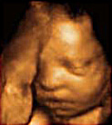
At Create Health we offer a comprehensive 2D, 3D and 4D (active real-time motion and expression) ultrasound scanning to all pregnant women, using the latest cutting edge technology. The vast majority of ultrasound scans are done through the abdomen – although in early pregnancy and when examining the cervix and low lying placenta at any stage, the transvaginal scanning is optimal.
We also offer a comprehensive non-invasive prenatal screening by biochemical and ultrasound screening program for congenital anomalies and chromosomal abnormalities at various stages of pregnancy.
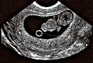
Ultrasound Diagnosis in the First Trimester
Assessment of gestational age by crown rump length. This is particularly important for any women who are unsure of their dates because of irregular periods, conception after recently stopping the pill or whilst on the pill, and conception whilst breast feeding.
Diagnosis of pregnancy from 1 week after the first missed period. It is possible to visualize the gestational sac inside the uterus at this gestation, and confirm an ongoing pregnancy.
Diagnosis of fetal viability from 5.5 – 6 weeks. It is possible to detect heartbeat even at this early stage (1.5 – 2 weeks after the first missed period), and allows confirmation of a live fetus. Whether the pregnancy is in the uterus, or an ectopic pregnancy can be accurately assessed.
Diagnosis of ectopic pregnancy. A potentially life threatening condition when the pregnancy occurs in the wrong place, early diagnosis of ectopic pregnancy can be made through ultrasound and biochemical tests. Early diagnosis facilitates medical treatment and avoids a surgical operation. Thus appropriate medical or surgical treatment can be planned to avoid danger.
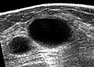
Diagnosis of multiple pregnancy and zygocity. Approximately 2% of women conceiving spontaneously and 10 -30% of women on fertility treatment will have a multiple pregnancy. At this stage of gestation it offers a unique opportunity to check if the fetuses share a common placenta, as this would have implications to for the rest of the pregnancy.
Screening for chromosomal abnormalities by nuchal translucency scanning between 11-14 weeks. By measuring the fluid behind the neck of the fetus at this stage of pregnancy, in combination with maternal age, we are able to give you a risk for major chromosomal abnormalities which is specific to you, You and your partner can then decide, based on the results, whether it’s high enough to warrant invasive prenatal diagnosis testing which can be chorionic villus sampling or amniocentesis later in the pregnancy.
Diagnosis of some major congenital abnormalities. Some of the major congenital abnormalities can be visualized at this early gestation, and allow for a plan of management to be decided upon.
Ultrasound Diagnosis in the Second Trimester
Fetal anomaly scan involving a detailed systematic survey of the fetus to detect congenital abnormalities and identify anatomical markers for chromosomal abnormalities. It is commonly done between the 18th to 22nd week of pregnancy. It allows major organs to be visualized and assessed as to its function and anatomy.
Doppler blood flow measurement : By measuring the blood flow in the uterine arteries at this stage of gestation, it is possible to pick out a sub-group of patients, which are at increased risk of developing pre-eclampsia and small babies. This allows treatment to commence at an early stage to modify the potential disease.
Placenta Location : A low-lying placenta, which covers the neck of the womb, may be a potential obstacle to a normal vaginal delivery. It may lead to serious bleeding later on in pregnancy endangering mother and baby. In cases where the placenta is visualized to be low at this gestation, we recommend that a scan should be repeated at 34th week, as most placental sites tend to move up and out of the way in late gestation as the uterus increases the size.
Diagnosis of Incompetent Cervix. By measuring the cervical length, it is possible to identify a group of patients who are at risk of premature labor, and it allows special management plan to be made for the pregnancy.
Ultrasound Diagnosis in the Third Trimester
Fetal Growth : The size and weight of the fetus can be determined by measuring a set of parameters. A scan is usually done around 32 - 33 weeks of gestation to screen for small gestational fetuses. Subsequent scans will be planned to monitor their growth.
Fetal Well-being : Ultrasound study of the behavior of the fetus constitutes the Biophysical Profile which is an indication of fetal well being. Together with Doppler Blood Flow we can find out if the fetus is happy and well inside the womb. It allows for an early detection of problems, and allows for treatment to minimize complications, including post delivery problems like cerebral palsy.
Identification of late fetal anomalies. Although relatively rare at this late stage to pick up any abnormalities in the fetus, it is possible that some fetal abnormalities might have been missed in early scans, or that certain conditions might have developed since the last scan.
Ultrasound Examination of The Breast
An ultrasound of the breast is performed when we need to 'look' for different abnormalities of the breast. For example, it is superior when trying to determine whether some lesion is cystic or solid.
By performing Doppler study, we may also be able to determine if there is any abnormal blood flow to the lesion, and aid in the diagnosis of lesions in the breast.
For some women this is required after a mammogram has shown a lesion which is suspicious, for others this maybe the preferred modality for examination.
Ultrasound guided biopsy can also be performed if indicated. Uniquely, The Women's Clinic can perform this under 4D guidance with our state of the art Kretz machine.